Mr Morritt regularly removes lumps such as lipomas (fatty lumps), sebaceous cysts (also known as epidermoid cysts) and nerve tumours (schwannomas). Lump removal surgery can generally be performed under local anaesthetic as a daycase procedure (go home the same day) for the majority of patients as the lumps tend to be relatively small. In the rare instances where lumps are either very large or are deeply located, a short general anaesthetic (patient is asleep) may be required.
Lipoma (fatty lump) removal
Mr Morritt frequently removes lipomas. These are common benign fatty lumps that grow slowly and tend to be painless. We do not know yet what causes lipomas although they do sometimes run in families suggesting a genetic cause. Lipomas can sometimes be painful (this is more common with ‘angiolipomas’). The vast majority of lipomas are small and are located immediately under the skin so can be moved around freely. These shallow lipomas can usually be removed through very small scars under local anaesthetic.
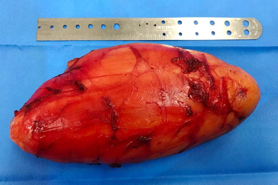
Lipomas can also be deeply sited both within muscles or underneath muscles. In certain locations or for large lipomas it is necessary for surgery to be performed under general or regional anaesthetic (such as spinal anaesthesia). Sometimes lipomas can grow to massive sizes e.g. the size of a watermelon. Mr Morritt has considerable experience in removing all lipomas and regularly undertakes surgery to remove lipomas from complex areas such as those located near major blood vessels or nerves.
Case 1. Photos showing appearances before and 6 weeks after removal of a large lipoma (fat lump) in the subcutaneous tissues of this lady’s tummy. Lumps like this grow slowly and stretch the skin so when they are removed it is often necessary to remove the excess skin. This lady had the lump removed under general anaesthetic (she was asleep for the procedure) because it was large and deeply located. Thanks to my patient for giving her permission to show these images.


Atypical lipomatous tumour (ALT)
A rarer fatty lump that can be mistaken for a lipoma is called an atypical lipomatous tumour (ALT or well differentiated liposarcoma). These are very similar to lipomas but regrow more frequently after they are removed. This diagnosis should be considered for all fatty lumps that measure greater than 5cm and are deeply sited. These lumps are a specialist area that is managed within the Sarcoma MDT. Mr Morritt is a core member of the sarcoma MDT and regularly removes these lumps.
Read more about removal of ALT/well differentiated liposarcoma

Dercum’s disease
Mr Morritt regularly treats patients with Dercum’s disease (multiple lipomas; also known as adiposis dolorosa). Sometimes these patients can have hundreds of lipomas. It is often not feasible to remove all the lipomas in the whole body from patient’s with Dercum’s disease as there are so many lumps. Mr Morritt therefore tends to recommend removal of any lumps that are painful, those that catch on clothing, those on cosmetically sensitive areas and those lipomas that are larger (typically greater than 5 cm diameter). Other lipomas can be removed for cosmetic reasons if patients desire.

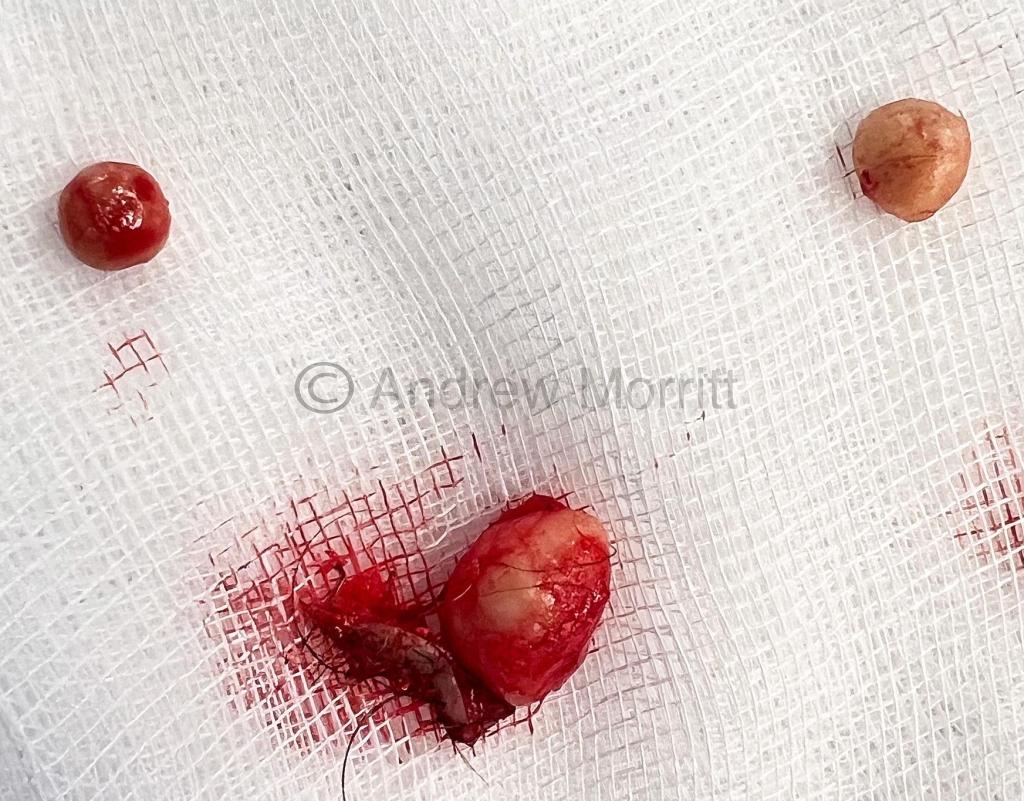
Case 1. Removal of 5 lipomas (fatty lumps) from the forearms of a patient with Dercum’s disease (multiple lipomatosis). Surgery was performed under local anaesthetic as a day case procedure. These lumps were angiolipomas which are a type of lipoma which can be painful. Thanks to my patient for giving consent to show these photos and for the lovely review.


Sebaceous cyst removal (Epidermoid cyst)
Mr Morritt frequently removes sebaceous cysts. These are cysts that occur in skin on areas of the body where there is hair. The typically range from the size of a pea to the size of a marble but can sometimes be much larger. Sebaceous cysts are very common and are benign. Rarely, if the cyst is subject to repeated inflammation or infection over many years, they can turn into a type of skin cancer called SCC (squamous cell carcinoma). Often patients may have squeezed the cysts thinking they are spots revealing the contents which are like cottage cheese and can smell. It is best not to squeeze these cysts because this makes them harder to remove surgically. Contrary to popular videos online where the cysts are lanced and their contents squeezed out, the best treatment is for the whole cyst to be removed. We know from experience that leaving the shell or part of the shell of the sebaceous cyst increases the chance of the cyst growing back.

Case 1. This gentleman in his 30’s had a sebaceous cyst excised from the left cheek. Appearances are shown before and 6 weeks after surgery. It is normal for the scar to be pink at this stage and with time the colour will change to match the normal skin colour more closely (this takes approximately 4-6 months in this location. In patients with darker skin the scar will initially become darker than the surrounding skin but this lightens with time (this is called post inflammatory hyperpigmentation). The surgery was performed under local anaesthetic (patient was awake) and stitches were removed 5 days after surgery. The cyst was removed with a small piece of skin and is shown in the image below. Thanks to my patient for giving permission to show these images.


Case 2. This gentleman in his 50s was troubled by some cysts on his scalp which although not being particularly painful got in the way. Appearances are shown before and 6 weeks after intact cyst removal. It is relatively common for there to be a slight indentation after removal of a lump or cyst but with time this and the scar becomes less noticeable.

Case 3. Removal of multiple epidermoid cysts from a patient’s scalp. Some patients have multiple sebaceous cysts and this can also run in families. Here the cysts have been removed under local anaesthetic – meaning that the patient was awake during surgery. Sometimes a small area of overlying skin is removed with the cyst. As you can see the cysts have been removed intact (not punctured) which is my preference as it reduces the potential for regrowth. Stitches are removed 12-14 days after surgery (self dissolving stitches are available for patients who do not want to have stitches removed after surgery).

Case 4. Before and after appearances following removal of a cyst from this gentleman’s chin. This cyst/lump was thought to be a sebaceous or epidermoid cyst. Normally these lumps are removed without taking any surrounding skin however having removed many cysts in the past, I noticed some subtle differences. I therefore removed the cyst with some surrounding skin (black ink marks on the before photo). The analysis showed that this was actually a rare cyst called a syringoma and not a sebaceous cyst. Thankfully it was the benign (non cancerous) version and as surrounding skin had been removed my patient did not need any further surgery. Thanks to my patient for giving permission to show these images.

Removal of nerve lumps
Mr Morritt regularly removes nerve lumps. These are most frequently benign lumps. They are often called peripheral nerve sheath tumours (PNST’s) or schwannomas. PNST’s arise from nerves and are generally benign although rarely can be malignant (cancerous). Nerve lumps are typically very tender and tapping them may result in electric shock type feelings. Sometimes PNSTs can arise on major nerves in the body and Mr Morritt is experienced in removing nerve lumps in that situation. Rarely complete removal of a nerve lump may result in the removal of part of or the whole nerve and in that situation Mr Morritt can undertake reconstructive surgery to rebuild the nerve with a nerve graft which is a piece of nerve taken from elsewhere on the body. A very rare condition exists called MPNST (malignant peripheral nerve sheath tumour) which is a cancerous nerve lump. MPNST typically requires major surgery and radiotherapy for treatment.
Another type of nerve lump is called a neurofibroma. These benign lumps are generally fleshy tags on the skin and can be removed with minor surgery. Some patients may suffer from a condition called neurofibromatosis where their whole body may be covered in neurofibromas. Rarely the neurofibroma may grow on a major nerve and removal typically requires reconstructive surgery to rebuild the nerve.
Review from patient who had nerve lump (schwannoma) removed by Mr Morritt

Lump removal – more patient reviews


